Ki-67
概要
KI-67は MKI67 遺伝子にコードされる、細胞分裂関係タンパク質である。ゆえに増殖細胞マーカーとして用いられており、臨床では肺がんに関わるマーカーとして利用されている。
構造
N-terminal forkhead-associated (FHA) domain; a protein phosphatase (PP)1 binding domain , 16 tandem repeats of 122 residues (in primates); C-terminal LR (leucine/arginine-rich) chromatin-binding domain[4]
機能
分裂細胞マーカーとしての利用

Ki-67は増殖マーカーである。 [7] 細胞増殖に強く関係している。間期の間、Ki-67は核で強く検出され、有糸分裂の際にクロモソームの表面へと大多数が再配置される。[8] Ki-67は細胞周期のすべての活性化フェイズ(G1, S, G2, M期)に存在しているが、静止状態の細胞 (G0期)には存在しない.[9] 細胞内のKi-67量は細胞分裂S期に急激に増えることが知られている。[10]. またKi-67は肺がんで高い増殖能を持つ患者サブタイプ (ER-positive 肺がんで、アジュバント化学療法が大きな効果をもつ)の同定に用いられる。[11] [12]
抗体ラベル
Ki-67 is an excellent marker to determine the growth fraction of a given cell population. The fraction of Ki-67-positive tumor cells (the Ki-67 labeling index) is often correlated with the clinical course of cancer. The best-studied examples in this context are carcinomas of the prostate, brain and the breast and nephroblastoma and neuroendocrine tumours. For these types of tumors, the prognostic value for survival and tumor recurrence have repeatedly been proven in uni- and multivariate analysis.
MIB-1
Ki-67 and MIB-1 monoclonal antibodies are directed against different epitopes of the same proliferation-related antigen. Ki-67 and MIB1 may be used on fixed sections.[13] MIB-1 is used in clinical applications to determine the Ki-67 labelling index. One of its primary advantages over the original Ki-67 antibody (and the reason why it has essentially supplanted the original antibody for clinical use) is that it can be used on formalin-fixed paraffin-embedded sections, after heat-mediated antigen retrieval (see next section below).
Ki-67抗体の起源
The Ki-67 protein was originally defined by the prototype monoclonal antibody Ki-67,[14] which was generated by immunizing mice with nuclei of the Hodgkin lymphoma cell line L428. The name is derived from the city of origin (Kiel, Germany) and the number of the original clone in the 96-well plate.
See also
- PCNA - Proliferating Cell Nuclear Antigen, expressed during the DNA synthesis.
Additional images
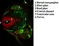 Immunofluorescent antibody staining against neurofilament (green) and Ki-67 (red) in a mouse embryo 12.5 days after fertilization. The proliferating cells are in the ventricular zone in the neural tube and therefore colored red.
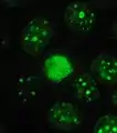 Protein Ki-67 in human MCF-7 cells.
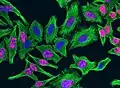 Ki-67 protein (red), tubulin (green) and DNA (blue) in HeLa cells. Dividing cells show strong Ki-67 staining in cell nuclei while all cells contain large amounts of tubulin, the major component of microtubules. Antibodies, cell staining and image courtesy of EnCor Biotechnology.
References
- “Entrez Gene: Antigen identified by monoclonal antibody Ki-67”. 2018年5月28日閲覧。
- “Assignment of the gene(s) involved in the expression of the proliferation-related Ki-67 antigen to human chromosome 10”. Hum. Genet. 83 (3): 297–9. (October 1989). doi:10.1007/BF00285178. PMID 2571566.
- “Ki-67 protein is associated with ribosomal RNA transcription in quiescent and proliferating cells”. J. Cell. Physiol. 206 (3): 624–35. (March 2006). doi:10.1002/jcp.20494. PMID 16206250.
- http://www.cell.com/trends/cell-biology/pdf/S0962-8924(17)30136-8.pdf
- https://www.nature.com/articles/nature18610
- “Chromophore-assisted light inactivation of pKi-67 leads to inhibition of ribosomal RNA synthesis”. Cell Prolif. 40 (3): 422–30. (June 2007). doi:10.1111/j.1365-2184.2007.00433.x. PMID 17531085.
- “The Ki-67 protein: from the known and the unknown”. Journal of Cellular Physiology 182 (3): 311–22. (March 2000). doi:10.1002/(SICI)1097-4652(200003)182:3<311::AID-JCP1>3.0.CO;2-9. PMID 10653597.
- “Ki-67 acts as a biological surfactant to disperse mitotic chromosomes”. Nature 535 (7611): 308-12. (July 2016). doi:10.1038/nature18610. PMC 4947524. PMID 27362226.
- “Cell cycle dependent expression and stability of the nuclear protein detected by Ki-67 antibody in HL-60 cells”. Cell Proliferation 25 (1): 31–40. (January 1992). doi:10.1111/j.1365-2184.1992.tb01435.x. PMID 1540682.
- “Initiation and termination of DNA replication during S phase in relation to cyclins D1, E and A, p21WAF1, Cdt1 and the p12 subunit of DNA polymerase δ revealed in individual cells by cytometry”. Oncotarget 6 (14): 11735–50. (May 2015). doi:10.18632/oncotarget.4149. PMC 4494901. PMID 26059433.
- “Final 10-year results of the Breast International Group 2-98 phase III trial and the role of Ki67 in predicting benefit of adjuvant docetaxel in patients with oestrogen receptor positive breast cancer”. European Journal of Cancer (Oxford, England : 1990) 51 (12): 1481–9. (2015). doi:10.1016/j.ejca.2015.03.018. PMID 26074397.
- “Ki67 in breast cancer: prognostic and predictive potential”. The Lancet. Oncology 11 (2): 174–83. (2010). doi:10.1016/S1470-2045(09)70262-1. PMID 20152769.
- Bánkfalvi A (November 2000). “Comparative methodological analysis of erbB-2/HER-2 gene dosage, chromosomal copy number and protein overexpression in breast carcinoma tissues for diagnostic use.”. Histopathology 37 (5): 411–9. doi:10.1046/j.1365-2559.2000.00984.x. PMID 11119122.
- “Production of a mouse monoclonal antibody reactive with a human nuclear antigen associated with cell proliferation”. Int. J. Cancer 31 (1): 13–20. (1983). doi:10.1002/ijc.2910310104. PMID 6339421.
- “Interaction of the chromatin compaction-inducing domain (LR domain) of Ki-67 antigen with HP1 proteins”. Genes to Cells : Devoted to Molecular & Cellular Mechanisms 7 (12): 1231–42. (2002). doi:10.1046/j.1365-2443.2002.00596.x. PMID 12485163.
参考文献
- Daniel G. Booth, and William C. Earnshaw. "Ki-67 and the Chromosome Periphery Compartment in Mitosis." Trends in Cell Biology. (2017) doi:10.1016/j.tcb.2017.08.001
External links
- Ki-67 Antigen - MeSH・アメリカ国立医学図書館・生命科学用語シソーラス(英語)
- http://www.pathologyoutlines.com/topic/stainski67.html
Template:PBB Controls